
 Re: GAMSAT Biology The Cell
Cell theory
The cell theory explains the observation that organisms are composed of cells. This theory includes the following three principles:
1. All organisms are made of one or more cells. Life processes occur in these cells.
2. Cells are the basic units of organisation in all organisms.
3. A cell can only arise by division of a previously existing cell.
Cell size
Cells are relatively small for a number of reasons related to diffusion in and out of the cell. A small cell is advantageous in terms of the surface area to volume ratio. A smaller, flatter cell with a large SA:V ratio will be more efficient in diffusion and removal of wastes. As a cell gets larger its volume increases at a faster rate than its surface area (lower SA:V ratio). A larger, rounder cell will have less efficient diffusion and removal of wastes.
Visualisation of cells
Many cells are not visible to the naked eye. In order to visualise the microscopic cells we need to use microscopy.
Types of microscopes
The aim of microscopy is to increase magnification so that cells can appear larger. Types of microscopes used in biology include:
Light microscope:
– Operate using visible light
– First lens focuses image onto second lens. Image is magnified and the image is focused on the back of the eye.
– Microscopes that magnify using multiple lenses are called compound microscopes.
– Able to resolve structures that are at least 200 nm apart.
Electrons have a much shorter wavelength and an electron microscope that uses electrons (instead of visible light) has 1000 x the resolving power than a light microscope.
Transmission electron microscope:
– Electrons transmitted through material
– Resolve images 0.2 nm apart
Scanning electron microscope:
– Beams electrons on the surface of the specimen. Electrons reflect back from the surface and other electrons from the specimen are released from the bombardment.
– Electrons are amplified and transmitted to a screen. A 3D image is produced.
Chemical stains can be used to increase the contrast between different components of the cell. Some structures either absorb or exclude the dye and this produces contrast that aids resolution.
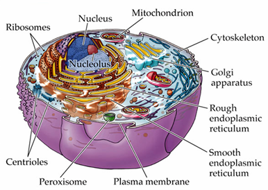
Cell structure
There are 4 major features that all cells have in common. These include:
Nucleoid or nucleus: Genetic material is located here.
Every cell contains DNA. Prokaryotes are the simplest organisms and contain a circular molecule of DNA. This DNA is found in the center of the cell in the region called the nucleoid. Eukaryotes are more complex organisms and their DNA is found in the nucleus. A double membrane called the nuclear envelope surrounds the nucleus.
Cytoplasm:
The cytoplasm is the semi-fluid matrix that fills the interior of the cell. The cytoplasm contains all of the amino acids, proteins and sugars that are essential to the cell.
Ribosomes:
Protein synthesis occurs at the ribosomes.
Plasma membrane:
The plasma membrane is a phospholipid bilayer with proteins embedded into it. The plasma membrane encloses the cell and separates the internal component of the cell with the environment.
Types of proteins in plasma membrane:
– Transport proteins: Help ions and molecules move across the plasma membrane. Movement occurs either in or out of the cell.
– Receptor proteins: Bring about changes within the cell when the receptors come in contact with molecules such as hormones.
Prokaryotes
Prokaryotes are small and are the simplest organisms. They have a plasma membrane that surrounds the cytoplasm. The plasma membrane is surrounded by a rigid cell wall. The rigid cell wall brings strength to the cell.
In prokaryotes the enzymes, DNA and other cellular constituents are not membrane-bound like they are in eukaryotes. Instead they have access to the entire interior of the cell.
The two main domains of prokaryotes are bacteria and archaea.
Bacteria: Bacterial cells have a cell wall that consists of peptidoglycan. The cell wall maintains the shape of the cell, protects the cell, and prevents excessive loss or uptake of water. Bacteria can also have an extra protective capsule that surrounds the cell wall.
Remember: Bacteria are often susceptible to antibiotics because the antibiotics affect the constitution of the cell wall.
Archaea: Archaea are difficult to culture and thus have not been studied in detail. Archaea have pseudopeptidoglycan in their cell wall.
Some prokaryotes have rotating flagella that enable movement. Flagella are protein fibers that extend from the cell and some cells may have one or more of these.
Eukaryotes
Eukaryotic cells are more complex than prokaryotic cells. They contain organelles that are membrane-bound structures that form compartments. Biochemical processes take place within each compartment.
Eukaryotes have a nucleus that contains DNA. DNA is wrapped into compact units called chromosomes within the nucleus. The nucleus is responsible for the synthesis of nearly all proteins in the living cell.
Most eukaryotes have one nucleus, but some fungi may have multiple nuclei. A nucleolus is a region in the nucleus in which synthesis of ribosomal RNA is taking place.
Chromosomes are composed of chromatin (DNA and protein complex). The DNA is wrapped around histones to form a nucleosome.
The nuclear envelope surrounds the surface of the nucleus in eukaryotes. It has two phospholipid bi-layer membranes. The nuclear envelope has pores that allow for the movement of proteins into the nucleus and for the exportation of RNA complexes from the nucleus to the cytoplasm.
Ribosomes are the cell’s protein synthesis machinery. Ribosomes translate mRNA to produce polypeptides. Polypeptides form proteins.
Eukaryotic cells have a cytoskeleton, which is an internal protein scaffold.
Fungi and plants have cell walls, whilst animals do not have cell walls.
Endomembrane system
The interior of a cell contains an endomembrane system. The role of this system is to allow for the channeling of molecules through the interior of the cell and provide surfaces for the synthesis of some proteins and lipids. This system contains rough and smooth endoplasmic reticulum (ER).
The rough endoplasmic reticulum is the site for protein synthesis. It is studded with ribosomes and synthesises and modifies proteins.
The smooth endoplasmic reticulum lacks ribosomes, but still has multiple roles. It is involved in lipid and carbohydrate synthesis.
The Golgi apparatus sorts and packages proteins. It receives vesicles from the ER and then modifies, repackages and transports them.
Lysosomes are also components of the endomembrane system in eukaryotes. They contain digestive enzymes that break down molecules and recycle the components of old organelles.
Mitochondria and chloroplasts
Mitochondria and chloroplasts contain their own DNA and have a double membrane structure.
The function of mitochondria is to metabolise sugar to generate ATP. The mitochondria have a highly folded inner membrane that contains proteins. These inner membrane proteins, along with the surface proteins, carry out metabolism to produce ATP.
Chloroplasts utilise light to generate ATP and sugars. The chloroplasts work by capturing light energy via thylakoid membranes arranged in stacks called grana. The light is essential in the process of forming glucose.
Cytoskeleton
The function of the cytoskeleton is to anchor the organelles and support the shape of the cell. It consists of crisscrossed protein fibers. The cytoskeleton can also help move materials within the cell.
Cell Movement
Flagella and cilia aid in the movement of the cell. Flagella arise from a basal body and cilia are shorter and more numerous than flagella.
Fungi
Fungi are eukaryotic organisms. They can be unicellular (yeast) or multicellular (filamentous fungi – mushrooms). Fungi can reproduce both sexually and asexually.
Virus
Viruses are very small obligate intracellular parasites. This means that in order to replicate their genetic material and thus multiply, they must be inside a living cell.
Viruses are considered non-living as they have no cellular structure and cannot carry out their metabolism independently.
Viruses may have DNA or RNA as their genetic material, but they cannot have both. The genetic material of a virus is found in their protein coat, which is termed the capsid.
On a final note, if you would like some free practice GAMSAT questions click the link below:
==> Free GAMSAT Mock Exam